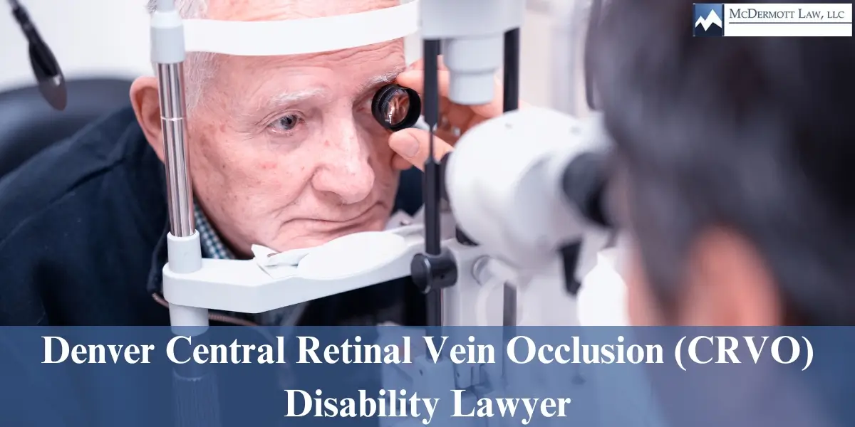
Denver Central Retinal Vein Occlusion (CRVO) Disability Lawyer
Denver Central Retinal Vein Occlusion (CRVO) Disability Attorney
Central Retina Vein Occlusion (CRVO) is a common retinal vascular disorder. Clinically, CRVO presents with variable visual loss; the fundus may show retinal hemorrhages, dilated tortuous retinal veins, cotton-wool spots, macular edema, and optic disc edema.
If you have been diagnosed with Central Retina Vein Occlusion (CRVO), it’s crucial to seek legal assistance from an attorney who specializes in CRVO disability cases. Contact our expert lawyers team at McDermott Law, LLC to discuss your case. Our experienced Denver CRVO Disability Lawyers are dedicated to helping you navigate the complexities of your claim and ensuring you receive the compensation you deserve.

Kooragayala, Lakshmana M., M.D., Central Retinal Vein Occlusion. Medscape (updated June 29, 2016), http://emedicine.medscape.com/article/1223746-overview
Cystoid Macular Edema (CME)
Cystoid macular edema (CME) is a painless disorder which affects the central retina or macula. CME occurs when fluid and protein deposits collect on or under the macula of the eye (a yellow central area of the retina) and causes it to thicken and swell (edema). The swelling may blur or distort a person’s central vision, as the macula is near the center of the retina at the back of the eyeball. This area holds tightly packed cones that provide sharp, clear central vision to enable a person to see detail, form, and color that is directly in the direction of gaze. Although the exact cause of CME is not known, it may accompany a variety of diseases such as retinal vein occlusion, uveitis, or diabetes. It most commonly occurs after cataract surgery
Cystoid Macular Edema (CME). Kellogg Eye Center (reviewed by Grant M. Comer, M.D., M.S.), https://www.umkelloggeye.org/conditions-treatments/cystoid-macular-edema-cme
Diabetic Retinopathy
Diabetic Retinopathy is caused by changes in the blood vessels of the retina. When these blood vessels are damaged, they may leak blood and grow fragile new vessels. When the nerve cells are damaged, vision is impaired. These changes can result in blurring of your vision, hemorrhage into your eye, or, if untreated, retinal detachment.
Diabetic retinopathy is the most common diabetic eye disease and a leading cause of blindness in the United States. Additionally, individuals with diabetic retinopathy may experience complications related to otolaryngology diseases, such as issues with hearing or balance, as diabetes can affect multiple systems in the body.
Macular Hole
A macular hole is a small break in the macula, located in the center of the eye’s light-sensitive tissue called the retina. The macula provides the sharp, central vision we need for reading, driving, and seeing fine detail. Macular holes often begin gradually. In the early stage of a macular hole, people may notice a slight distortion or blurriness in their straight-ahead vision. Straight lines or objects can begin to look bent or wavy. Reading and performing other routine tasks with the affected eye become difficult. The size of the hole and its location on the retina determine how much it will affect a person’s vision. When a Stage III macular hole develops, most central and detailed vision can be lost. If left untreated, a macular hole can lead to a detached retina, a sight-threatening condition that should receive immediate medical attention.
Facts About Macular Hole. National Eye Institute (May 22, 2018),
Optic Neuritis
Optic neuritis is an inflammation that damages the optic nerve, a bundle of nerve fibers that transmits visual information from your eye to your brain. Pain and temporary vision loss in one eye are common symptoms of optic neuritis.
Optic neuritis is linked to multiple sclerosis (MS), a disease that causes inflammation and damage to nerves in your brain and spinal cord. Signs and symptoms of optic neuritis can be the first indication of multiple sclerosis, or they can occur later in the course of MS. Besides MS, optic neuritis can occur with other infections or immune diseases, such as lupus.
Symptoms include pain, vision loss in one eye, visual field loss, loss of color vision, and seeing flashing lights associated with eye movement.
Optic Neuritis. Mayo Clinic. https://www.mayoclinic.org/diseases-conditions/optic-neuritis/symptoms-causes/syc-20354953
Oscillopsia
Oscillopsia is a visual disturbance in which objects in the visual field appear to oscillate. The severity of the effect may range from a mild blurring to rapid and periodic jumping. Oscillopsia is an incapacitating condition experienced by many patients with neurological disorders.
http://en.wikipedia.org/wiki/Oscillopsia
Post Trauma Vision Syndrome
A person who has suffered a TBI may often experience difficulties with balance, spatial orientation, coordination, cognitive function, and speech. Usually a referral for visual consultation only occurs if there’s an injury to an eye or if ocular pathology is suspected. Persons with a TBI frequently experience symptoms of double vision, movement of print or stationary objects (such as walls and floor), eye strain, visual fatigue, headaches, and problems with balance. Visual problems are among the most common sequalae following a TBI, but frequently not dealt with in a rehabilitation model.
Retinal Detachment
The retina is the light-sensitive layer of tissue that lines the inside of the eye and sends visual messages through the optic nerve to the brain. When the retina detaches, it is lifted or pulled from its normal position. In some cases there may be small areas of the retina that are torn. These areas, called retinal tears or retinal breaks, can lead to retinal detachment. Symptoms include a sudden or gradual increase in the number of floaters, which are little “cobwebs” or specks that float about in your field of vision, and light flashes in the eye. Another symptom is the appearance of a curtain over the field of vision. A retinal detachment is a medical emergency, and if not promptly treated, can cause permanent vision loss.
Facts About Retinal Detachment. National Eye Institute (May 22, 2018), https://nei.nih.gov/health/retinaldetach/retinaldetach
Uveitis
Uveitis is the swelling and irritation of the uvea, or the middle layer of the eye which provides most of the blood supply to the retina. It can be caused by autoimmune disorders like rheumatoid arthritis, infection, or exposure to toxins, but in many cases the cause is unknown. The most common form is anterior uveitis which involves inflammation in the front part of the eye. This is called iritis, as it involves the iris. Posterior uveitis affects the back part of the eye and involves the choroid which is a layer of blood vessels and connective tissue in the middle part of the eye. Individuals may develop this condition following a body-wide (systemic) infection or an autoimmune disease. Symptoms of uveitis can include blurred vision, dark or floating spots in the vision, eye pain, redness of the eye, and sensitivity to light.
Facts About Uveitis. National Eye Institute (May 22, 2018),
Visual Tracking Issues
To use our vision efficiently, the eyes must move accurately, smoothly, and quickly from lace to place. Eye movements allow accurate scanning of the visual environment for information. Every time a person looks from afar to something near them, , their eyes must accurately jump from one target to another. The same is true for reading as the eyes jump from one word to another. Tracking is also important for following moving objects in sports, and for directing our eyes to move our hands towards a target. Eye-hand coordination in any activity starts with accurate eye movements. Tracking skills are considered the fine motor aspect of vision, and like other fine motor skills, should continue to develop into adulthood. With tracking problems, eye movements are slow, inaccurate, or require head or finger movement to help the eyes track. This can interfere with reading fluency and comprehension, copying, handwriting, and sports performance. Those with tracking problems may show the following behaviors or signs: frequent loss of place during reading, skipping lines, omitting and substituting words during reading, head movement or finger pointing during reading, slow reading speed, poor copying, and poor eye-hand coordination. In Denver, where factors like altitude and environmental conditions may also contribute to visual processing challenges, these symptoms can sometimes be exacerbated.
www.drgallaway.com/vision-and-learning/eye-teaming-focusing-and-tracking
Contact Us
For more information about your legal rights and entitlements, along with a professional evaluation of your potential case, set up a free Review of Denied Claims by calling us at 303-964-1800, or complete our contact form. We offer free Review of Denied Claims and represent clients throughout the Denver area and the Rocky Mountain region.
Get A Free Case Evaluation of Your Denied Claim
Fields Marked With An “*” Are Required


